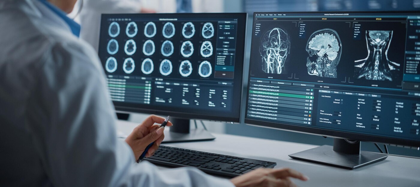
Unruptured brain aneurysms -- bulging areas in the brain’s blood vessels -- can be dangerous and even deadly if they leak or rupture. In the past, determining whether to observe the unruptured brain aneurysm or operate has mostly hinged on the aneurysm’s size and location. But that paints an incomplete picture, says David Hasan, MD, a Duke Health neurosurgeon and researcher who has been working for decades to improve brain aneurysm diagnosis and treatment. He and his colleagues at Duke are using sophisticated tools to help you determine the best next steps.
Here are some of the innovative ways Duke uncovers information about your unruptured brain aneurysm and how to treat it.
Advanced Imaging for Brain Aneurysms
One of the most important factors in deciding how to treat your unruptured brain aneurysm is its stability. An unstable brain aneurysm has a higher risk of rupturing and causing a stroke (doctors call this a subarachnoid hemorrhage), and that can have devastating effects. According to Dr. Hasan, these imaging options, available for select patients at Duke, are groundbreaking for determining brain aneurysm stability.
Molecular Imaging with Ferumoxytol
Ferumoxytol is an injectable medication used to treat people with chronic anemia. Before coming to Duke, Dr. Hasan and his team discovered1 that the ferumoxytol molecule acts like a contrast agent when used with MRI imaging to detect specific inflammatory cells responsible for aneurysm eruption.
“Think of it like a volcano,” Dr. Hasan said. “If there are a lot of inflammatory cells present, it's like the volcano is rumbling and ready to erupt. On the other hand, if you don't have many inflammatory cells, the aneurysm is quiet and cool.”
Because of the side effects associated with ferumoxytol, it’s not right for everyone. It can be used in people with kidney problems because it’s easier on the kidneys than traditional MRI contrast.
High-Resolution Vessel Wall Imaging
This technique uses traditional MRI contrast called gadolinium. By suppressing a specific MRI signal, the contrast pools in the area between the inner wall of the brain aneurysm and the blood inside. A white color indicates that the aneurysm has active changes, increasing the risk of rupture (whereas a black color indicates the opposite).
Quantitative Susceptibility Mapping (QSM)
QSM is a specific setting or sequence that is added to an MRI scan to help detect and measure micro-bleeds -- when trace amounts of blood leak through very thin areas of the aneurysm wall. These micro-bleeds can signal that an aneurysm rupture is coming.
QSM is especially useful in people with an unruptured brain aneurysm and migraines or tension headaches, since severe headache is a hallmark symptom of aneurysm rupture.
Computational Dynamics
This method uses MRI and computer modeling to measure how fast blood moves through the aneurysm. Slower blood movement is associated with aneurysm deterioration.
Finite Element Analysis
Finite element analysis uses MRI technology to assess the strength and elasticity of the brain aneurysm wall. A weaker and less elastic aneurysm wall is more likely to rupture.
Photon-Counting CT
While coil embolization is the most common treatment option for unruptured and ruptured intracranial aneurysms because it’s highly effective and minimally invasive, a major disadvantage of coil embolization is that up to 20% of people experience aneurysm recurrence. We developed a dedicated 3D imaging process that detects recurrence by analyzing changes to the coil using a new platform of non-contrasted computed tomography (NCCT) of the brain called photon-counting CT. Unlike more traditional angiography methods, this method does not require contrast dye to be injected, which is safer, more economical, and more convenient for patients.
Research Opportunities, Innovative Procedures Available at Duke
Dr. Hasan and his colleagues are also working to move brain aneurysm care forward in other ways, like studying devices to record activity in brain arteries and researching aspirin use to treat unruptured brain aneurysms. In addition, Duke is one of a handful of centers in the U.S. offering “awake surgery” for endovascular surgical procedures that treat brain aneurysms.
“Duke is unique because we have advanced research protocols, and patients want to have access to these opportunities,” Dr. Hasan said. “They want to be part of the research, even if it doesn't help them at this point. Maybe it will help others in the future.”
Dr. Hasan, an international authority in cerebrovascular care and research, has treated thousands of brain aneurysms using innovative techniques, including awake surgery.


