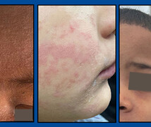
Parents frequently ask questions about strawberry marks -- called hemangiomas -- in their infants and children Will they stay? Will they grow? Will they go away?
Jane Bellet, MD, a pediatric dermatologist at Duke Health, offers information and advice to address these common questions about hemangiomas.
Hemangiomas Are the Most Common Birthmark
The most common birthmark is a hemangioma. One in every ten babies has one, yet the cause is still unknown. A hemangioma usually develops during the first two weeks after birth, often as a small, red, flat area or bump. This type of hemangioma is called “superficial” and involves the surface of the skin. The bump can continue to grow for the next nine to 12 months, when it begins to slowly involute -- or shrink and fade in color.
Some hemangiomas are subcutaneous, which means they are below the surface of the skin and often appear blue. Many hemangiomas have both superficial and subcutaneous components, so they appear red on top and blue underneath.
The majority of hemangiomas will never cause a problem and do not require treatment because their appearance will gradually diminish with time. Since hemangiomas grow most rapidly between five weeks and six months of age, it is important that infants be evaluated early, as treatments are often most effective during the growth phase.
Hemangioma Treatments
Children with hemangiomas that require treatment should be referred to a pediatric dermatologist. The location of the birthmark is one of the most important considerations because most hemangiomas located on or near the eyelid, nose, lip, and beard area -- the jawline or in front of the ear -- need treatment to prevent complications. These may include vision loss, feeding problems, and respiratory distress. Hemangiomas in the beard area may be associated with airway involvement.
Large or ulcerated hemangiomas -- when a sore or wound develops on the skin over the hemangioma -- also require treatment. Infants with more than five hemangiomas may also have them in other locations such as the liver or gastrointestinal tract, and these can grow just as hemangioma on the skin do. Appropriate imaging should be performed to determine if treatment is necessary.
A large plaque-like hemangioma on the face indicate PHACE syndrome:
Posterior fossa malformations
Hemangioma-segmental
Arterial abnormalities of the neck or brain
Cardiac-often coarctation of the aorta
Eye abnormalities
Treatment is determined for each individual child as a number of factors are involved. Historically, hemangiomas were treated with oral corticosteroids such as prednisolone. Doctors now know that propranolol is more effective, and it is now the first-line treatment when systemic treatment is needed. Fortunately, propranolol also has fewer severe side effects compared to oral corticosteroids. Topical timolol is also used in some situations. Ulcerated hemangiomas require a multi-faceted approach including pain management and treatment of infection, if present. Once hemangiomas have stopped growing and have entered the involutional phase, any remaining redness can often be addressed with laser therapy. Excisional surgery may be required to remove any extra skin.
Prognosis
In most cases, hemangiomas are not problematic and can be easily diagnosed and observed by your child’s pediatrician. When treatment is required, early referral and management by a pediatric dermatologist who specializes in hemangiomas is highly recommended.


