If salivary stones are causing you pain or other annoying symptoms, Duke Health offers a minimally invasive procedure called salivary endoscopy (also called sialendoscopy). Instead of traditional, more invasive surgery to remove the affected gland, salivary endoscopy requires specially trained head and neck surgeons using delicate tools to remove the stones, resulting in a much quicker and easier recovery.
What Are Salivary Stones?
Also called sialolithiasis, salivary stones are calcified mineral deposits that get stuck in the glands that produce saliva or the ducts that transport saliva to the mouth. Common symptoms include pain and swelling while eating, a lump under the tongue, bad breath, dry mouth, and bitter-tasting or gritty saliva. Left untreated, salivary stones can cause tooth decay, infection, and even breathing problems.
Doctors review CT scans or ultrasound imaging to determine the size, location, and shape of the salivary stone(s) when deciding whether sialendoscopy is your best treatment option. They also consider whether your duct or gland is infected and any other health concerns that could play a role.
What Is Sialendoscopy?
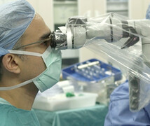
Developed in the 1980s, sialendoscopy requires specialized surgical training and tiny, fragile, and expensive fiberoptic endoscopes. “You’re talking about something that is the size of the tip of a ballpoint pen.” Duke Health’s Liana Puscas, MD, MHS, said. “They require extra gentle handling, and you need to have several in reserve.”
Traditionally, large salivary stones are treated by surgically removing the obstructed gland. This leaves a scar, requires a lengthy recovery, and runs the risk of damaging nearby facial nerves. In contrast, during sialendoscopy, a surgeon uses tiny tools to widen the opening of the affected salivary duct. Then a tiny metal basket traps and retrieves the stone. Finally, a few stitches are added to ensure the duct stays open so saliva can flow freely. It takes about 90 minutes from start to finish and doesn’t require a hospital stay.
Follow-Up and Beyond
After the procedure, your mouth will be sore for a few days, and you’ll need to eat only soft foods, according to Dr. Puscas. Once the swelling goes down, people usually notice a difference right away. A follow-up visit can be done in person or through a telehealth visit.